
Having a child is a great experience, probably an exciting one too. But it is also very confusing and sometimes even frustrating when you are expecting for the first time and have little to no idea how things work. A common question that parents have when having their first child is ‘how to read ultrasound numbers,’ and that is what we will handle here.
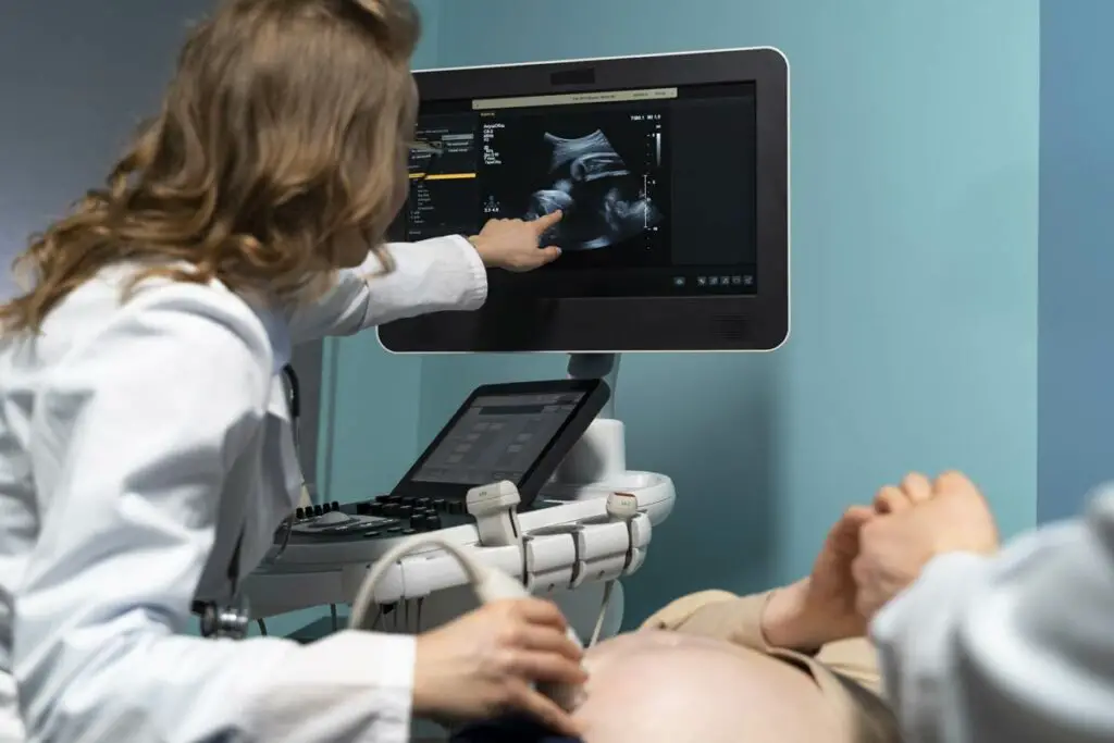
Many pregnant women are given an ultrasound (sometimes known as a sonogram) as prenatal screening. It displays an image on a monitor or gives out a printed image of the baby inside the uterus (womb) using sound waves. Ultrasound images can assist in monitoring normal prenatal development and screening for potential abnormalities. Along with a regular ultrasound, there is also advanced medical imaging, such as a 3-D ultrasound, a 4-D ultrasound, and fetal echocardiography, a type of ultrasound that examines the fetus’ heartbeat in depth.
The CM label on the pregnancy ultrasound shows the gestational age of the baby in weeks. It means ‘Mean Sac Diameter. This metric is commonly described as the length in cm from the pelvic bone to the roof of the womb. After week 24, the fundal height (CM) of a properly developing baby is expected to correspond to the number of weeks pregnant — with an additional or lesser 2 cm in certain cases. Prenatal ultrasound measurements might include parietal diameter, head circumference, crown length, abdominal circumference, etc.
For those parents who are seeing their very first ultrasound images, it could be hard to understand all the different numbers and images. The scan might look confusing at first, but all those measurements provide more details about your child. The most common issue many new parents face is interpreting the numbers on the ultrasound images.
Ultrasound Numbers
Typically, the top of an ultrasound picture displays a sequence of numbers as well as other information. This area is commonly used by ultrasound clinics and hospitals for information such as:
- Name of the patient
- Hospital identification number
- Ultrasound machine settings
Those numbers and information also assist you in identifying the front of ultrasound pictures. But this isn’t always the case. The upper section of an ultrasound picture is determined by the location of the ultrasound probe. In the image, you should be able to spot a cone-like object. The ultrasound image expands from a tiny to a huge portion. The upper half of the ultrasound picture is at the smaller end.
Information on an Ultrasound Picture
Because they’re non-invasive and give real-time pictures, pregnancy ultrasound scans are a common way of testing and diagnosis.
Ultrasound machines produce high-frequency sound waves. These waves are directed toward the body and enter the skin. The sound waves are then bounced against a person’s tissue or internal organs.
The bounced ultrasound waves are recorded by ultrasound equipment; therefore, the resultant patterns create images of a person’s inside tissues and internal organs. Ultrasound interpretations provide information on the state of the body’s soft tissue, major organs, or fluids.
Abbreviation on an Ultrasound Picture
There are several abbreviations you may encounter during an ultrasound of the developing baby. Sonographers and OB-GYNs often utilize these to monitor a baby’s development as well as discover any irregularities. This works to ensure that every aspect of the pregnancy goes as planned.
You may come across the following ultrasound abbreviations:
AC – Abdominal circumference; the millimeter measurement of an abdomen, one of four fundamental measurements used to determine a baby’s growth or gestational age.
BPD – Biparietal diameter; determines the diameter of the skull of the baby, another fundamental metric used in conjunction with AC, FL, and HC to determine age or weight.
CPR – Cerebroplacental ratio; often used to indicate unfavorable outcomes such as placental instability in future pregnancies.
CRL – Crown-rump length; a measure of the baby from the crown of the head to just under the tailbone, used to calculate gestational age.
EDD – Estimated date of delivery; an expected date for the delivery depending on the size or age of the fetus.
EFW – estimated fetal weight; calculated baby weight based on fundamental measurements
FHM – Fetal heart movements; the monitoring of the fetus’ pulse.
FL – Femur length; another primary measurement made by the sonographer; it’s the size of the femur.
GA – Gestational age; it’s your baby’s estimated age relying on the previous menstrual cycle and measurements.
GS – Gestational sac; among the primary ultrasound readings that could be taken, measures the sac or fluid surrounding the fetus.
LMP – Last menstrual period; starting date of the last menstrual period, often used to determine the conception date and expected due date.
HC – Head circumference; this measures the baby’s head in millimeters, a fundamental measurement used along with BPD, FL, and AC.
MSD: Mean gestational sac diameter; a measure of the gestational sac used to determine the aging process.
SGA – Small for gestational age; it’s used when the baby is smaller than the expected size for the pregnancy stage.
TVS – Trans V scan; the pelvic ultrasound performed through the V canal that often detects early pregnancy.
Transverse Spine, Umbilical Vein, and Stomach
The fetal ultrasound images of the transverse spine, umbilical vein, and stomach are among the most important images, due to the importance of these organs:
Transverse plane of the spine:
Any potential anomaly in the spine must be scanned in the transverse plane to establish the existence of splaying. It’s critical to photograph perpendicular to every vertebra in the transverse plane of the spine.
When scanning the transverse plane of the spine, the curving of the fetal spine must be considered to prevent utilizing too much craniocaudal angulation, which might produce the illusion of splaying, or “pseudodysnaphism.”
Umbilical vein:
The umbilical vein (UV) transports oxygenated blood from the placenta to the developing baby. The umbilical vein (UV), portal vein (PV), and ductus venosus (DV) are all essential components of a developing fetus. UV and DV screening should be done as part of a standard second-trimester anomaly scan.
Umbilical Vein Varix (focal dilatation of the UV), PRUV (Right Umbilical vein devolves during the first 4-6 embryonic weeks while the left persists), and DV agenesis (a drain that predominantly sends highly oxygenated blood to the left heart) can all be identified.
Stomach ultrasound:
A stomach ultrasound is a traditional ultrasound diagnostic used to monitor the health of a pregnant woman. This examination is frequently referred to as a prenatal ultrasound by practitioners.
Stomach ultrasonography may also aid in determining the source of unexplained abdominal (stomach) pain. This test assists in the identification of numerous common illnesses (such as stone formation) as well as more significant health issues (like blood clots).
First, Second and Third Trimester Baby
First trimester:
During the first 90 days, most ultrasound images are used to examine an embryo’s development, verify the number of babies, monitor the amniotic fluid, determine the age of the fetus, and predict the due date of delivery (EDD).
Second trimester:
Ultrasounds conducted between weeks 18 to 20 enable an ultrasound technician to monitor fetal growth and take fetal measurements of specific features such as appendages, spinal column, internal organs, the baby’s skull and brain. If any parents wish to know, they can also verify the size and positioning of the placenta, the baby’s age, as well as the gender.
Third trimester:
Ultrasound images beyond week 30 or day 210 of a pregnancy can be used to evaluate the baby’s average ongoing growth. The placement of the amniotic fluid can also be checked to verify that it is not obstructing the cervix.
Crown-rump Length
Throughout pregnancy, ultrasounds are used to assess the crown-rump length (CRL). The baby’s height is calculated in cm from the crown of its head to just the end of its hips (rump). The measurement excludes the limbs or yolk sacs. The CRL may be measured at six and seven weeks into the pregnancy and up to 14 weeks.
CRL might be useful in determining the baby’s age. Doctors can predict the due date based on that. The sooner the baby’s age is determined, the more precise it’s going to be.
Diameter of the Baby
Biparietal diameter (BPD) is one of many parameters measured during prenatal ultrasound scans. It refers to the diameter of a maturing baby’s skull measured through one parietal bone to the second. The baby’s weight, as well as gestational age, are calculated using biparietal diameter.
Most people receive one to three ultrasounds from early in pregnancy until around week 20. People who are deemed to face a significant threat may require further ultrasounds.
A BPD measurement is beneficial in conjunction with three additional measurements:
- Circumference of the baby’s head
- The circumference of the abdomen
- Length of the femur bone (the longest bone in the body and is located in the thigh)
These three measurements, when combined, serve in estimating fetal mass as well as gestational age. The BPD assessment also provides you and your doctor with information about how your growing baby’s central nervous system is progressing.
Your doctor is aiming for the BPD and other parameters to be within the healthy range. At 13 weeks, the biparietal diameter measurement is around 2.4 centimeters; at term, it’s about 9.5 centimeters.
How to Read Ultrasound Images for Gender?
The ultrasound reading for gender is usually performed halfway through the pregnancy. The prenatal anatomy survey is performed to search for potentially fatal illnesses rather than merely determining your baby’s gender.
The reliability of the ultrasound imaging will be determined by a variety of factors, including the infant’s age, the equipment utilized, the ultrasound technicians and technique, the ultrasound centers as well as the baby.
What’s the Difference Between a Girl and Boy Ultrasound Scan?
Signs of a Female Ultrasound
When using ultrasound images to determine the gender of a fetus, the sonographer will search for distinguishing traits known as signals. There are two indications to look for in a girl:
Hamburger sign: The shape of the female anatomy and cervix on ultrasound is referred to as a “Hamburger sign.” If one looks carefully at the ultrasound image, one will see that the V lips form a hamburger bun, whereas your adult female letter “C” area will resemble a hamburger patty on an ultrasound.
Sagittal sign: There is a sagittal sign for each sex. It is determined by examining the fetus’s profile (the midline sagittal plane). The caudal notch, a bump, may be found near the end of the backbone. It’s a female if it is pointed downward at a 10-degree inclination.
Signs of a Boy Ultrasound
By weeks 18 to 20, a newborn male would be determined based on the following signs:
Sagittal sign: If the caudal notch is pointed upwards towards an angle greater than 30 degrees, the fetus is a male. It becomes more difficult to reach a firm conclusion if it falls somewhere in the middle.
Urine flow: A fetus’s urine stream can be seen on occasion. If it is rising upward, it’s more likely to be a male.
Male genitalia: Often visible between weeks 18 and 20, the existence of the male genital area on the ultrasound image, including the gonads, balls, or obvious male genitalia, is a definite indication of the male sex.
Other Methods of Determining Sex
A chorionic villus sampling (CVS) or amniocentesis can be used in parallel to an ultrasound to determine the gender of your unborn child. Amniocentesis is a technique that involves extracting fluid from the sac that surrounds your baby using a syringe and a needle. CVS entails extracting cells from the placenta. Both these ways are highly accurate methods of determining the gender of the baby.
Be Prepared
During pregnancy, you should do your best to be prepared for the experience of being a parent. The pregnancy period is a time for you to learn as much as you can about how you can give your child the best life. The ultrasound images can help you determine whether your child is healthy or needs additional medical attention. Most importantly, you will understand how your child is developing, and that is a gift on its own.
